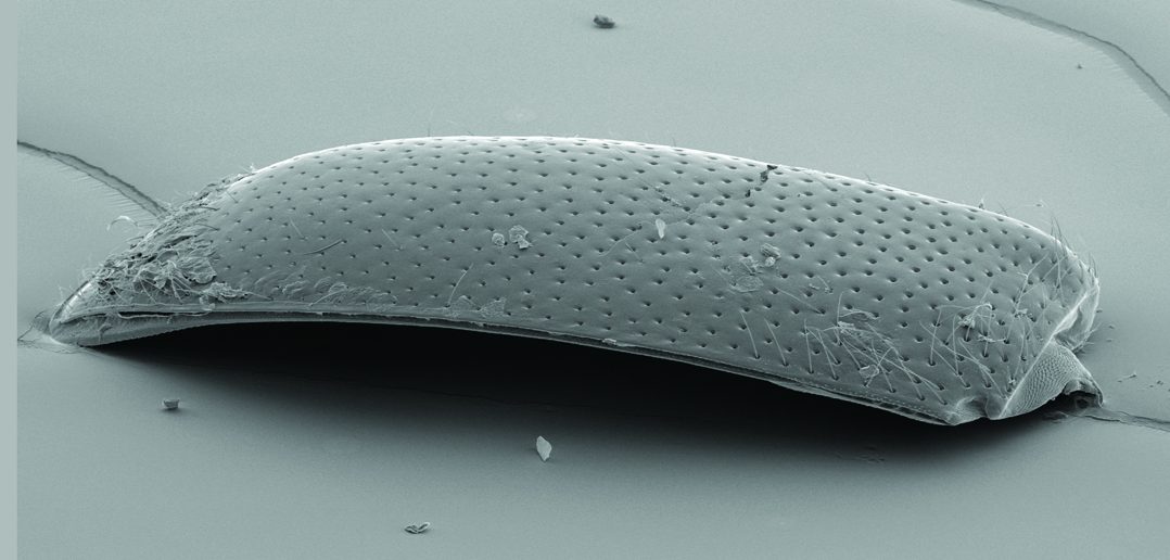
Writ large.
Microscopist and photographer Geoff Williams, MS, finds artistic opportunity in unusual places. His daughter’s eye, for example. Five years ago, when she was 6, a beetle collided with her face while she was playing soccer, lodging a chitinous forewing under her eyelid.
“She had to go to Hasbro [Children’s Hospital] and be sedated because the pediatric ophthalmologist couldn’t get close enough with forceps to pluck it off without her flinching,” Williams says. “The shape was really cool,” so he took the 1 mm-long wing back to Brown’s Leduc Bioimaging Facility, coated it with a film of pure gold, and put it in a scanning electron microscope. At nearly 350x magnification, it’s even cooler: you can see her corneal cells stuck to the wing.
When Williams displays his work, he doesn’t always identify the object. “The point is trying to get people to first visually, aesthetically react, and then maybe want to figure out what they’re looking at,” he says.
See the prepared beetle wing specimen and read more about Williams in Anatomy of a Microscopist.


