In ways old and new, visuals play a key role in how we learn, teach, and practice medicine.
In the Blue Room of Hasbro Children’s Hospital, Danielle Sawka ’22 MD’26 stands off to the side as six figures in blue huddle over the operating table. On it, a tiny shape draped in sheets is bathed from above in bright white light, a Betadine-swabbed belly poking through an opening in the fabric. A herd of machines on wheels hisses and beeps.
Like a conductor raising his baton, the hospital’s pediatric surgeon-in-chief, François Luks, MD, PhD, initiates the timeout that is part of the Universal Protocol: “Baby girl Prentiss. Laparoscopic gastrostomy.” The anesthetist, then the circulating nurse confirm the name of the patient and the procedure. Luks points to the mark where the incision will be. “Are there any concerns?” There are none. The operation begins.
Wasting no time, Sawka steps closer to the table. Only her watchful, wide-open eyes, the shade of a pale winter sky, are visible between the surgical cap and mask. Three monitors suspended from the ceiling by giant tentacles suddenly come to life as a telescope—a thin, wand-like tube containing a camera and a light cable—descends a cannula that’s been inserted in the baby’s abdomen. The camera moves forward gingerly, projecting a view of liver, colon, and stomach. Low-ceilinged and pink, the baby’s insides look like the GoPro footage of a spelunker in a Himalayan salt cave. Pincers come into view and grip the veiny flesh of the stomach. “You see how we tent the stomach, Danielle, so the guidewires can be inserted and anchor the G-tube?” Luks says.
Sawka’s gaze locks onto one screen and then another, then drops to the large sketch pad cradled in her left arm. With a pencil she makes a few tentative dashes on the paper, looks up again, and delivers a flurry of marks. A series of sketches with hasty annotations cascades down the paper, marking the steps of the procedure. She turns the page quickly and keeps drawing as she tries to keep up with the surgeon.
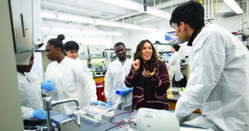
As a student in Luks’s popular course, Introduction to Medical Illustration (PLME 0400), Sawka is here not to do the surgery or even just watch it, but to see it. What began in 2010 as a two-hour workshop for first-year medical students is now a semester-long course offered to Brown and Rhode Island School of Design undergraduates that covers not only the fundamentals of drawing, such as point of view, light and shadow, outline and contour, but the history and ethics of medical illustration as well. In addition to live models and plant and animal specimens at RISD, students sketch cadavers in the anatomy lab and operations like this one. They hear from practicing medical illustrators, such as Ian Suk, from the Johns Hopkins School of Medicine’s storied Department of Art as Applied to Medicine, as well as Julia Lerner MD’23, who trained there, and the Warren Alpert Medical School’s own Vinald Francis, hired last summer.
Medical drawings have existed for more than 2,000 years, evolving from third-century BC renderings on papyrus, to Leonardo da Vinci’s 15th-century Vitruvian Man, to Andreas Vesalius’s 16th-century De Humani Corporis Fabrica, to the works of 20th-century stars Max Brödel and Frank Netter. Today’s artists have replaced sable brushes and carbon dust with styluses and software like Photoshop, but their role, according to the Association of Medical Illustrators, is the same: they are visual problem solvers.
Luks, who is also professor of surgery, of obstetrics and gynecology, and of pediatrics, is quick to point out that the goal of his course is not to turn students into Leonardos, but to teach them to communicate—with their patients, with students, with each other. “If you convert big words into easy language, it’s still very abstract. If you draw it, it’s concrete,” he says. “An image sticks.” Lerner considers drawing a particularly important tool to use with patients, “especially when there are barriers in health literacy, or even language.”
It’s also a great teaching tool for oneself, Luks says, since to be effective, even the most rudimentary drawing requires a thorough understanding of the pathology or procedure: “Think about a complicated operation. How do you attack it? You break it down in its simplest parts, then figure out what’s essential and what is not.” To do this, Luks has his students watch a video of a gallbladder removal several times, and then represent it—in only three drawings.
In addition to this analytical thinking, medical drawing demands close observation, which Francis points out is an integral part of medical training. “By the time you’re actually [doing]a procedure, you’ve seen it many, many times. And you understand it inside and out. … No amount of lecturing can take the place of that.”
Sawka adds that while such close observation is important, just as useful is “the ability to filter out certain details” to make the visual appropriate to the viewer’s level of understanding. Both skills are key for clinicians, as Luks, Kevin Liou ’10 MD’15, Paul George ’01 MD’05 RES’08, and Jay Baruch, MD, director of the Program in Clinical Arts and Humanities, pointed out in a 2014 paper in Medical Education: “The ability to select which details are relevant to the larger picture … is also essential in non-visual aspects of clinical practice, such as in formulating diagnoses and delivering oral presentations.”
Sawka says that “the chance to learn from such an accomplished surgeon, medical illustrator, and professor” as Luks makes PLME 0400 the best course she’s ever taken. It’s now being replicated by Jill K. Gregory, associate director of instructional design at Icahn School of Medicine at Mount Sinai.

This drawing of the ophthalmic artery by Vinald Francis, a medical illustrator at Brown, appeared in an article in the journal Stroke in February.
LESS IS MORE
This paring-down to the pith has its most extreme expression in what’s known as the visual abstract, in which the key points of a scientific paper are represented in a three-panel format using simple, stylized icons accompanied by succinct text—and then tweeted. The first visual abstract is attributed to a surgical resident at the University of Michigan, Andrew Ibrahim, who in July 2016 tweeted an infographic on a yellow background linking to “The Impact of a Pan-regional Inclusive Trauma System on Quality of Care in London,” which appeared in the Annals of Surgery. Since then, the trend has been consecrated with a hashtag and endorsed in a tweet by Atul Gawande (“Love the #VisualAbstract format. More journals need to pick this up.”).
And it has spawned a revolution in how scientific information is shared. To date, some 80 journals and organizations have integrated visual abstracts into their dissemination strategy; some even require authors to submit a visual abstract and tweet along with their paper. (Stroke was the first, in 2017.) The yellow-and-black design has become the Annals of Surgery’s signature look, and Ibrahim, now the journal’s creative director, has published an open-access online primer on the topic. Meanwhile, in June 2019, an editorial in the Journal of the American Geriatrics Society compared a text-only tweet to a tweet with a visual abstract; the authors found that the standard tweet received 24,984 impressions, 17 retweets, and 36 likes over eight days, while the post with the visual abstract received 168,447 impressions, 81 retweets, and 100 likes in half that time.
While visual abstracts have been compared to movie trailers or previews, Luks calls this approach the “IKEA model” of nonverbal communication. “Nobody’s better than IKEA at describing something visually without words in as few images as possible. They’ve turned it into an art.” Luks, who enjoys creating visual abstracts and tweets, sits on the social media committee of the American Pediatric Surgical Association; has spoken at SciVizNYC, a multidisciplinary gathering on the visualization of scientific information; and presented twice at the Association of Medical Illustrators conference.
Given the convergence of several social phenomena—the overwhelming amount of material doctors need to consume to stay up to date, the increasing prevalence of user-focused design thinking, the use of smartphone technology—not to mention scientific journals’ basic mission to disseminate research, it’s perhaps not surprising that health care professionals prefer this concise, DIY approach. The science shows that the brevity of visual abstracts might be easier for working memory to process, and graphics are more likely to be encoded in long-term memory, making them easier to retain and recall.
The downside to visual abstracts is that so far, they’re neither shareable beyond social media nor searchable on PubMed. But Luks refutes the criticism that visual abstracts constitute a dumbing down: “Most of my colleagues draw, some better than others. But we all use images. It’s what we’ve always done.”
THE PICTURE OF HEALTH
Visual language works because “no one speaks jargon,” Luks says. Technical medical terms can impede effective communication—especially at critical times in a patient’s care. Sheyla Medina MD’20, who all her life has used drawing to learn concepts, has seen the role art can play in compassionate, patient-centered care.
During a gap year as a Bray Fellow in Medical Humanities, Medina worked at Women & Infants Hospital’s Women’s Intimacy and Sexual Health Clinic with patients who had been treated for gynecologic and breast cancers. She says that explaining concepts through images rather than words enabled the women to “process and digest some of the hidden meanings behind their curiosities about treatment effects on the body.”
Medina wondered if there was a way to combine visual art with effective storytelling through comics. She partnered with Luks, who also had been thinking about medical comics, and Fred Schiffman, MD, the Sigal Family Professor of Humanistic Medicine, to secure an Arnold P. Gold Foundation grant on the use of visual communication—as opposed to the more commonly used essay—as a tool of reflection and expression. Despite the rise of narrative medicine and the popularity of journaling, Medina says, “not everyone processes with words.”

A panel from “The Palliative Path,” a medical comic by Frank Deng ’20.
For their pilot project, Medina, Luks, and Schiffman recruited seven undergraduates or recent graduates (including one RISD student) who had completed PLME 0400. Each student had to convey, through a comic, their conception of what humanism in medicine means. Over the course of five interactive workshops, they learned the technical basics and narrative tricks of comics with the help of Emily Slapin, an instructor at RISD, and received feedback on their work. The comics were displayed in the Medical School last fall.
The workshop leaders found that students were highly motivated to integrate humanities into their careers and considered comics to be a valuable way to communicate concepts and facilitate reflection. Shortly after the exhibition, Medina traveled to Orlando to present the findings at the Gold Humanism Society’s inaugural Gold Humanism Summit. Several of the comics will be published in the Annals of Internal Medicine.
Medina, who was born in Peru, explored meanings of health and well-being across cultures as a public health major at the University of Pennsylvania. Years before she was drawing medical comics, she observed the healing power of storytelling when she interviewed Ojibwe in Minnesota about their experiences with western and traditional medicine for a research project. One of her conclusions: “The stories themselves are medicine.”


