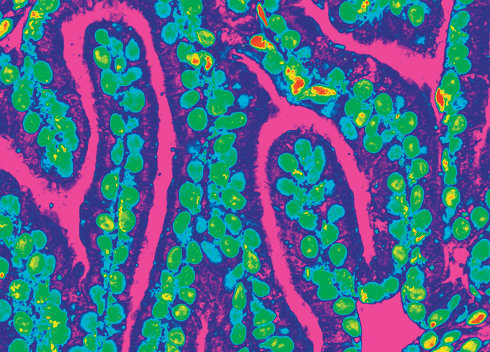
Under the microscope, science meets art.
For Harvey J. Kliman, MD, PhD, the microscope is a tool of the scientist and the artist. “Only in a Woman,” the first art exhibit mounted in the Alpert Medical School building, which ran through January, was composed of 16 immunohistograms of the endometrium and uterine structures that Kliman magnified and edited.
The piece shown here, “Pink Rivers,” is a false-color image of closely packed endometrial glands. With filters and further digital processing, Kliman transforms raw microscopic images into brilliant expressions of color and form.
Kliman is a research scientist in the Department of Obstetrics and Gynecology at the Yale University School of Medicine and the director of the Reproductive and Placental Research Unit, where his research interests include infertility and pregnancy complications. He studies the clinical utility of abnormalities in placental villous growth patterns to diagnose genetic abnormalities in pregnancy, including autism.
Lucy Partman, the exhibit’s curator, writes that “Kliman’s artwork provides an opportunity to explore the connections between art and science in an effort to see what they can reveal. How do you see and experience these images? How does scientific knowledge affect your perception?”


