Fine focus.
Childhood seemed pretty idyllic for Geoff Williams, MS. Sure, the Seattle native was a “struggling musician’s kid,” but he always had a camera, played the violin with his family, and rode his bike everywhere. “We grew up on a dead end road, and we had a hill,” he says. “We’d ride down the hill and crash at the bottom.”
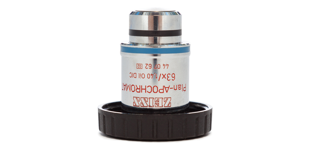
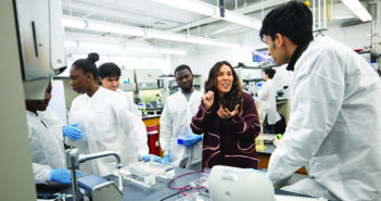
He’s still riding (and only rarely crashing) his bike, logging thousands of miles year-round on his daily commute; on punishingly long routes around southern New England; and racing cyclocross, a steeplechase-type, mostly off-road (and usually muddy) event that mixes pedaling, running, and obstacle jumping at relentless speeds. It’s a high-octane contrast to his day job in the sedate, dark labs of Brown’s Leduc Bioimaging Facility, where, as the manager, he keeps a dozen microscopes up and running and teaches students and faculty how to use them.
For Williams, microscopy labs are more than a place to indulge his mechanical mind or, as a botanist, to observe the minutest details of the plant world; they’re also an art studio. Whenever he has a few minutes to spare, he zooms in on everyday objects—bike parts, insects, kitchen scraps—to explore their aesthetic possibilities; he prints his favorite images and occasionally displays them in local galleries and exhibits. Examining pollen and other hidden treasures of a tiny juniper specimen in a scanning electron microscope (SEM), he says, “It’s a different world inside here that you don’t see everywhere else.”
Click here to see the magnified image of a beetle wing.


